An Introduction to Live-Cell Super-Resolution Imaging
Siân Culley, Pedro M Pereira, Romain F Laine, Ricardo Henriques (see publication in Journal )Abstract
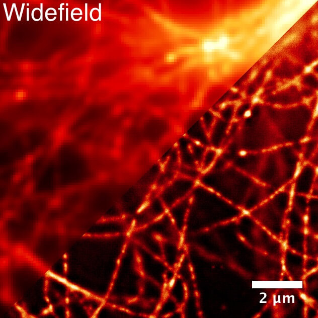
Fluorescence microscopy has been a crucial tool in the advancement of modern cell biology because of its nonin-vasive nature, compatibility with imaging live samples, and molecule-specific labeling tools. However, the resolving power of conventional fluorescence microscopy is limited to~ 250–300 nm. To resolve cellular structures on a smaller size scale than this, researchers have typically relied on electron microscopy, which can provide insight into structures on a nanometer scale. While electron microscopy continues to be a valuable tool for investigating fine intracellular structures, incompatibility with live samples and its limited labeling capabilities remain obstacles to studying dynamic phenomena with high confidence in molecular identities. This has led to the development of super-resolution microscopy methods, which were developed in the early 2000s. Super-resolution microscopy bridges the resolution gap between conventional fluorescence microscopy and electron microscopy while retaining the advantages associated with light microscopy. This chapter provides a brief overview of commonly used super-resolution microscopy techniques, their applications to live-cell imaging, and future directions for this family of techniques.