Live Imaging of a Hyperthermophilic Archaeon Reveals Distinct Roles for Two ESCRT-III Homologs in Ensuring a Robust and Symmetric Division
Andre Arashiro Pulschen, Delyan R Mutavchiev, Siân Culley, Kim Nadine Sebastian, Jacques Roubinet, Marc Roubinet, Gabriel Tarrason Risa, Marleen van Wolferen, Chantal Roubinet, Uwe Schmidt, Gautam Dey, Sonja-Verena Albers, Ricardo Henriques, Buzz Baum (see publication in Journal )Abstract
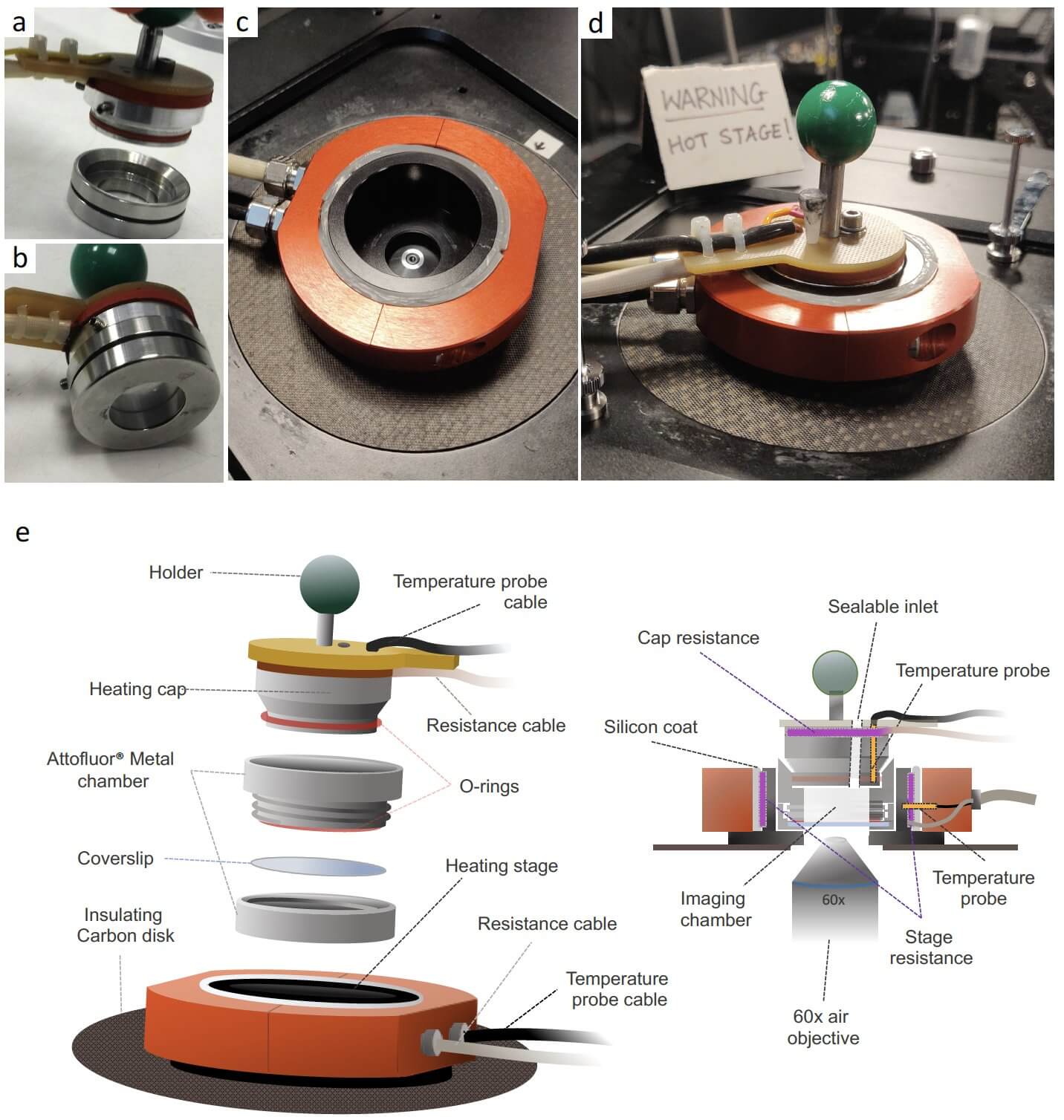
Live-cell imaging has revolutionized our understanding of dynamic cellular processes in bacteria and eukaryotes. While similar techniques have recently been applied to the study of halophilic archaea, our ability to explore the cell biology of thermophilic archaea is limited, due to the technical challenges of imaging at high temperatures. Here, we report the construction of the Sulfoscope, a heated chamber that enables live-cell imaging on an inverted fluorescent microscope. Using this system combined with thermostable fluorescent probes, we were able to image Sulfolobus cells as they divide, revealing a tight coupling between changes in DNA compaction, segregation and cytokinesis. By imaging deletion mutants, we observe important differences in the function of the two ESCRTIII proteins recently implicated in cytokinesis. The loss of CdvB1 compromises cell division, causing occasional division failures and fusion of the two daughter cells, whereas the deletion of cdvB2 leads to a profound loss of division symmetry, generating daughter cells that vary widely in size and eventually generating ghost cells. These data indicate that DNA separation and cytokinesis are coordinated in Sulfolobus, as is the case in eukaryotes, and that two contractile ESCRTIII polymers perform distinct roles to ensure that Sulfolobus cells undergo a robust and symmetrical division. Taken together, the Sulfoscope has shown to provide a controlled high temperature environment, in which cell biology of Sulfolobus can be studied in unprecedent details.