Nuclear pores as versatile reference standards for quantitative superresolution microscopy
Jervis Vermal Thevathasan, Maurice Kahnwald, Konstanty Cieśliński, Philipp Hoess, Sudheer Kumar Peneti, Manuel Reitberger, Daniel Heid, Krishna Chaitanya Kasuba, Sarah Janice Hoerner, Yiming Li, Yu-Le Wu, Markus Mund, Ulf Matti, Pedro Matos Pereira, Ricardo Henriques, Bianca Nijmeijer, Moritz Kueblbeck, Vilma Jimenez Sabinina, Jan Ellenberg, Jonas Ries (see publication in Journal )Abstract
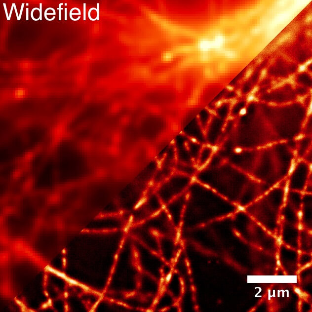
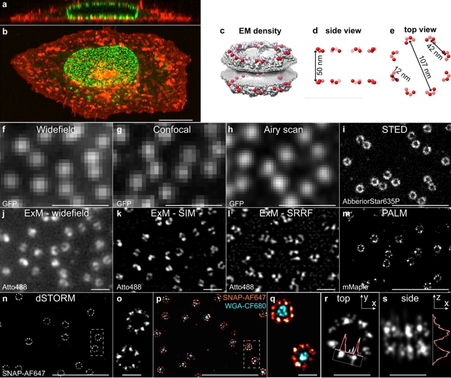
Quantitative fluorescence and superresolution microscopy are often limited by insufficient data quality or artifacts. In this context, it is essential to have biologically relevant control samples to benchmark and optimize the quality of microscopes, labels and imaging conditions. Here, we exploit the stereotypic arrangement of proteins in the nuclear pore complex as in situ reference structures to characterize the performance of a variety of microscopy modalities. We created four genome edited cell lines in which we endogenously labeled the nucleoporin Nup96 with mEGFP, SNAP-tag, HaloTag or the photoconvertible fluorescent protein mMaple. We demonstrate their use (1) as three-dimensional resolution standards for calibration and quality control, (2) to quantify absolute labeling efficiencies and (3) as precise reference standards for molecular counting. These cell lines will enable the broader community to assess the quality of their microscopes and labels, and to perform quantitative, absolute measurements.